Article · 16 October 2026
Enzyme Activity Assays in Rare Disease Drug Programs: Design, Validation, and Interpretation Across Four Clinical Contexts
A single enzyme activity assay format cannot equally serve diagnosis, pharmacokinetics, pharmacodynamics, and gene therapy efficacy readouts in lysosomal storage disorder programs. A 2026 review in Bioanalysis and supporting primary literature clarify how assay purpose, matrix, and immunogenicity monitoring must be aligned before validation begins. This article is for Research Use Only.
AlpinaBioTech

The Core Problem: One Analyte, Four Different Questions
A bioanalytical scientist supporting a lysosomal storage disorder (LSD) program may be asked to measure enzyme activity in the same patient sample at four distinct decision points: to confirm diagnosis, to characterize baseline pharmacokinetics of an enzyme replacement therapy (ERT), to read a pharmacodynamic response, and to assess whether a gene therapy construct has delivered functional protein. Each question looks identical on the surface, but the assay requirements, the acceptable bias, and the consequences of a wrong answer differ considerably at each stage.
A review article by Catherine Vrentas and colleagues, published in Bioanalysis in October 2026, addresses exactly this challenge [1]. The paper, "Applications of enzyme activity assays in clinical development for rare disease therapeutics," arrives at a moment when the number of ERT, gene therapy, and cell therapy programs targeting inborn errors of metabolism is expanding, and when bioanalytical scientists are being asked to design assays that must serve multiple regulatory and clinical functions at once.
Why Rare Disease Programs Stress the Assay
The diseases targeted by these programs are individually rare but collectively significant. The benchmark Australian incidence figure comes from Meikle et al. (JAMA, 1999), which studied 27 different LSDs diagnosed across Australia from January 1980 through December 1996: as a group of disorders, the combined prevalence was 1 per 7,700 live births [2]. A more recent Australian study covering the period 2009 to 2020 found a combined prevalence of 1 per 4,800 live births, considerably higher than the earlier estimate, partly reflecting improved ascertainment and a broader diagnostic case mix [3]. Other sources note that this figure may be as high as 1 in 5,000 births when all LSDs are considered [4]. The variation in these estimates reflects both the breadth of the disease category and the challenge of ascertainment in small patient populations, both of which have direct implications for clinical trial design and biomarker strategy.
The current standard of care for enzyme deficiencies is ERT and/or small molecule chaperones, with in vivo gene therapy and ex vivo cell therapy as promising new therapeutic approaches for the treatment of hereditary monogenic disorders [5]. To support the development of these different therapeutics, assays to quantify the functional activity of protein enzymes have gained importance in diagnosis, assessment of pharmacokinetics and pharmacodynamic response, and evaluation of drug efficacy [5]. A single assay format is unlikely to satisfy all four of those demands equally well. Fluorometric assays offer sensitivity well-suited to low-abundance targets in cerebrospinal fluid or dried blood spots, but they may lack the specificity to distinguish endogenous residual activity from recombinant enzyme in a patient receiving ERT.
The Gene Therapy Layer
Gene therapy programs introduce a specific bioanalytical complication that ERT programs do not face in the same way: the enzyme activity readout is no longer a direct measure of the drug itself, but a functional consequence of gene expression in transduced cells.
For AAV-based gene therapy products, the transgene protein is the product of the gene therapy molecule, and enzyme activity shows the functional effect after the vector infects or transduces the cell [1]. This makes the connection between transgene protein expression and enzyme activity critical for evaluating biodistribution and pharmacodynamics. The matrix from which enzyme activity is measured determines what biological question is actually being answered. For example, IDUA activity from dried blood spots and from white blood cells has been used as a readout in MPS I gene therapy studies using both lentiviral ex vivo and AAV in vivo approaches [6]. The choice of matrix is not merely logistical. It determines whether the assay is a PK readout, a pharmacodynamic readout, or a biodistribution surrogate. Conflating these will produce data that are individually correct but collectively uninterpretable at the clinical level.
Different gene therapy strategies are under clinical investigation in several LSDs, and enzyme activity biomarkers can be reliable substitutes for clinical responses and improve the efficiency of clinical trials [6]. This is the context in which enzyme activity assays function not as simple descriptors of enzyme presence, but as instruments that must be precisely calibrated to answer a specific regulatory or clinical question.
Complicating matters further, many patients enrolled in gene therapy trials are already on ERT at baseline. The assay must be capable of distinguishing residual signal from prior ERT from the emerging activity driven by the gene construct, particularly in the early weeks after dosing [1].
Go / No-Go Criteria: Where Assay Design Meets Clinical Decision-Making
One of the most concrete contributions of validated enzyme activity assays in rare disease programs is their role in prospective decision thresholds. The Vrentas et al. review describes a strategy in which clear criteria are set to determine whether the observed transgene protein expression and enzyme activity from gene therapy treatment is sufficient to achieve a clinically meaningful reduction of accumulated substrate [1].
This framing shifts enzyme activity from a descriptive biomarker to a decision-support tool. It requires the assay to be validated not only for precision and accuracy in the analytical sense, but also for its ability to detect differences that are biologically and clinically meaningful. A coefficient of variation that is acceptable for a diagnostic assay may be entirely inadequate for an assay expected to distinguish a therapeutic response from background variability.
Enzymatic activity plays a critical role throughout the drug lifecycle, influencing patient selection, gene therapy target engagement, PK and PD readouts, immunogenicity assessment, and surrogate endpoints [1]. Each of these applications carries a different tolerance for uncertainty, and the assay must be designed with that tolerance explicitly defined before validation begins.
Validation: Where the Field Currently Stands
The validation framework for enzyme activity assays does not map cleanly onto standard ligand binding assay (LBA) or LC-MS/MS guidance. A 2024 white paper-themed article by Azadeh, Vrentas, Quadrini, and colleagues in The AAPS Journal (Volume 26, Issue 5) addressed this directly, discussing the technical aspects of enzyme activity assays in the bioanalytical context, including assay design and format as well as the unique challenges and considerations associated with assay development, validation, and lifecycle management [5].
This paper is a review with white paper-style recommendations, not a primary methods study. Its authorship spans multiple industry and academic groups, consistent with its consensus function. The Quadrini and Vrentas GM1-gangliosidosis assay data, which the 2024 AAPS article cites, appears in a separate primary research publication: Quadrini et al. validated high-sensitivity fluorometric assays to quantify beta-galactosidase activity in cerebrospinal fluid and serum from patients with GM1-gangliosidosis, published in Molecular Therapy: Methods and Clinical Development in August 2024 [7]. That CSF assay is described as the first of its kind with sufficient sensitivity to quantitatively measure beta-galactosidase enzyme activity in CSF samples from GM1 patients [7].
The enzymatic assay development lifecycle closely mirrors a standard bioanalytical workflow, but with potentially increased importance placed on critical reagent management and lifecycle maintenance [5]. This matters practically because the substrate, the cofactor, and any reference enzyme standard used in the assay can degrade or shift between lot numbers in ways that alter the apparent activity reading without any change in the patient sample.
A parallel challenge applies across other LSD programs. For enzyme defects, biomarkers assessable from patient serum are highly useful, but activity assays need to be properly validated to ensure a precise, quantitative measurement [8]. Banning et al. established and validated a fluorometric AGA activity assay for human serum samples from healthy donors and AGU patients, demonstrating it is suitable for diagnostics and, potentially, for following a treatment effect [8]. The fit-for-purpose principle applies here as it does in LBA validation, but the definition of "fit" is more context-dependent than in small-molecule PK assays. The 2024 AAPS article recommends using LBA biomarker method validation as a general principle with a fit-for-purpose approach for certain steps, while remaining mindful of potential instability for some analytes and methods [5].
Immunogenicity: The Readout That Can Nullify Yours
Any scientist running enzyme activity assays in an ERT or gene therapy program should be tracking immunogenicity data in parallel. Patients on ERTs may develop anti-drug antibodies (ADA), which can be either neutralizing, directly inhibiting enzyme activity, or non-neutralizing, accelerating enzyme clearance.
This phenomenon is best characterized in Pompe disease. Administration of recombinant human GAA (rhGAA) triggers an immune response in some patients, particularly those with two null GAA variants that cause complete absence of detectable endogenous GAA on Western blot, who are characterized as cross-reactive immunological material (CRIM) negative [9]. About two-thirds of patients with classic infantile Pompe disease produce inactive GAA protein and are CRIM-positive; one-third produce no detectable GAA protein and are CRIM-negative [10].
CRIM-negative patients produce no detectable endogenous GAA protein and develop sustained high antibody titers to ERT that render the treatment ineffective [11]. Pharmacokinetic studies of ERT-treated patients with infantile Pompe disease have demonstrated an average increase in clearance of infused rhGAA by 50% from week 1 to week 12 in patients with high rhGAA IgG antibody titers at week 12, with antibody-mediated reduction or diversion of active rhGAA from muscle tissues likely accounting for this therapeutic failure [9]. The immune response mechanism in infantile Pompe disease is an established and well-documented example; whether the same pattern applies to other ERT programs depends on the specific disease, therapeutic protein, and patient population, and should not be assumed to generalize without disease-specific evidence.
This creates a direct confound for enzyme activity interpretation. If a patient's measured activity is declining over successive study visits, the cause may be sub-therapeutic dosing, poor biodistribution, or the emergence of neutralizing ADA that is actively suppressing the response in vivo. The enzyme activity readout alone cannot distinguish between these scenarios. Running enzyme activity and ADA assays from matched longitudinal samples is the only way to interpret activity trends with confidence.
Practical Design Considerations
Several pre-analytical and analytical factors are particularly consequential for enzyme activity assays in this context.
Substrate concentration and Km. The assay must be run at substrate concentrations that allow detection across the full dynamic range expected in the study, from near-zero activity in severely affected patients to supra-physiological activity following effective gene correction.
Matrix effects. Activity in serum, plasma, dried blood spots, white blood cells, and CSF will not be directly comparable. Each matrix must be validated independently if data from multiple matrices are to be reported [1]. The Quadrini et al. GM1 work demonstrated this concretely: rapid processing and freezing of samples post-collection was critical to preserve enzyme activity, and the effects of hemolysis and blood contamination on beta-galactosidase activities in serum and CSF were characterized explicitly [7].
Endogenous activity. Unlike many LBA analytes, the endogenous form of the target enzyme is present in all samples, including controls. Assay selectivity must be established against this endogenous background, and the lower limit of quantitation must be anchored to clinically meaningful differences from baseline [8].
Stability. Enzyme activity is inherently time- and temperature-dependent. The stability of the substrate-enzyme complex under real-world sample handling conditions must be characterized as part of validation, not assumed [5].
Critical reagents. The source and lot qualification of the reference enzyme standard is the single point of greatest fragility in most validated enzyme activity assays. Lot-to-lot variability in a fluorogenic substrate can shift the apparent activity of an unchanged reference standard by amounts that would be clinically significant if undetected [5].
Summary
The October 2026 Vrentas et al. review in Bioanalysis reflects a field that has moved well past asking whether enzyme activity assays work, and toward the harder question of how they should be designed, validated, and interpreted differently depending on the therapeutic modality they are intended to support [1]. For ERT programs, the assay is primarily a PK and PD tool. For gene therapy programs, it is simultaneously a biodistribution readout, a target engagement measure, and a decision threshold instrument [1]. The immunogenicity layer, particularly the risk of neutralizing ADA in patients producing no detectable endogenous enzyme protein, overlaps both program types and must be monitored in parallel [9, 11]. Scientists who treat these as the same assay problem will generate data that are analytically sound but clinically ambiguous.
This article is for Research Use Only. The assays and frameworks described herein have not been reviewed for diagnostic use.
Sources
- [1] tandfonline.com
- [2] pubmed.ncbi.nlm.nih.gov
- [3] researchgate.net
- [4] sciencedirect.com
- [5] link.springer.com
- [6] cell.com
- [7] pmc.ncbi.nlm.nih.gov
- [8] ncbi.nlm.nih.gov
- [9] insight.jci.org
- [10] jpeds.com
- [11] pmc.ncbi.nlm.nih.gov
More articles
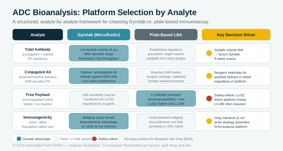
Platform Choice at the Core of ADC Bioanalysis: What the Gyrolab-vs-Plate Debate Actually Requires Scientists to Decide
Selecting between Gyrolab microfluidic and conventional plate-based immunoassay platforms for antibody-drug conjugate bioanalysis is not a single decision but a structured one made analyte by analyte. A sponsored interview published October 13, 2026, on Bioanalysis Zone, combined with the July 2026 AAPS ADC Working Group white paper in AAPS Journal, provides the clearest current framework for making those choices across total antibody, conjugated antibody, free payload, and immunogenicity assays. All assay content discussed is for Research Use Only.
Read more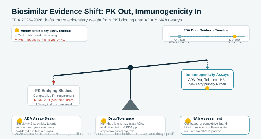
When PK Studies Slim Down, Immunogenicity Assays Carry More Weight: What FDA's 2026 Biosimilar Guidance Means for Bioanalytical Scientists
FDA's March 2026 draft guidance removes the default requirement for comparative PK bridging studies in biosimilar development, building on a prior October 2025 draft that proposed eliminating routine efficacy trials. Both actions shift evidentiary weight onto immunogenicity assays, raising the bar for ADA assay design, drug tolerance, and neutralizing antibody assessment before any clinical samples are collected.
Read more