Article · 21 September 2026
Immunogenicity Assessment for LNP-Based Therapeutics: Mapping the Assay Demands Across Innate and Adaptive Endpoints
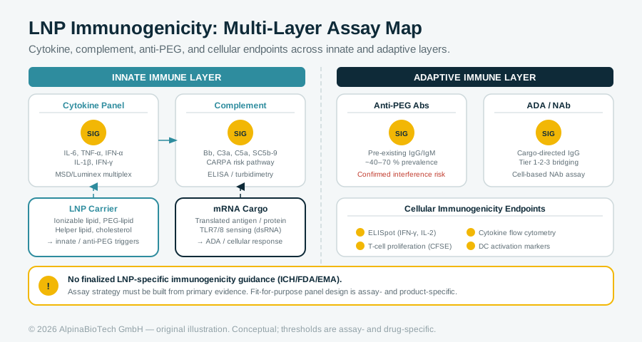
LNP-based therapeutics generate immunogenicity signals across at least two immune layers, involving carrier and cargo independently, against a background of pre-existing anti-PEG antibodies in a substantial fraction of patients. No finalized LNP-specific immunogenicity guidance exists, leaving bioanalytical scientists to build assay strategies from the LNP-specific evidence base up. This article maps the cytokine, complement, anti-PEG, and cellular endpoints that a fit-for-purpose panel must address.
AlpinaBioTech
For Research Use Only. Not for use in diagnostic procedures.
Immunogenicity Assessment for LNP-Based Therapeutics: Mapping the Assay Demands Across Innate and Adaptive Endpoints
Lipid nanoparticle (LNP)-based therapeutics are no longer a niche modality. Following FDA approvals of several LNP-based therapeutics and vaccines, including Onpattro (2018), Comirnaty (2021), Spikevax (2022), and mRESVIA (2024), understanding immune responses elicited by LNP-based products has become increasingly important. That clinical footprint creates a concrete problem for bioanalytical scientists: the immunogenicity signal from an LNP product is not a single, tractable readout. It spans at least two immune layers, involves the carrier and the cargo independently, and carries pre-existing background noise from environmental exposure in a substantial fraction of patients. Designing an assay strategy that is both scientifically justified and regulatory-ready requires working through each layer systematically.
Why LNP Immunogenicity Is Structurally Different from Protein-Therapeutic Immunogenicity
For a conventional monoclonal antibody or biosimilar, the immunogenicity question centers on whether the drug generates anti-drug antibodies (ADAs) that affect clearance or neutralize function. With LNP-based products, the question expands to cover at least three independent sources: the carrier components, the nucleic acid cargo, and, where the payload encodes a protein, the expressed protein itself.
In the context of LNP-based products, both LNP components and the encapsulated payload, such as mRNA, can be sensed by the innate immune system through pattern-recognition pathways, thereby activating innate immune responses. Moreover, when the payload encodes a protein, as in the case of mRNA, the resulting protein may elicit antigen-specific adaptive immune responses, including humoral and cellular immune responses [1].
The consequences of these immune responses may be beneficial or detrimental depending on the therapeutic indication and the magnitude, quality, and durability of the response [2].
The Regulatory Framework: ICH S6(R1), ICH S8, and the Absence of LNP-Specific Guidance
From a regulatory perspective, both the FDA and the ICH generally support a risk-based, product-specific approach to evaluating immunogenicity and immunotoxicity rather than requiring a predefined panel of immune assays for all LNP-based products [1]. ICH S6(R1) provides the preclinical safety framework for biotechnology-derived products, including consideration of humoral and cell-mediated immune responses. ICH S8 is more specifically scoped: it provides recommendations on nonclinical testing approaches to identify compounds that have the potential to be immunotoxic and guidance on a weight-of-evidence decision-making approach for immunotoxicity testing, restricted to unintended immunosuppression and immunoenhancement, and excluding allergenicity or drug-specific autoimmunity.
No LNP-specific immunogenicity testing guidance has been finalized. As confirmed by multiple independent bioanalytical commentaries, there is no clear regulatory guidance on this topic for LNP-mRNA products specifically [2, 13]. Practitioners are therefore working within the existing protein-therapeutic architecture, applied with scientific justification tailored to the LNP platform. Immunogenicity remains the principal constraint on the efficacy, safety, and re-dosing of LNP-mRNA therapeutics, and clinical decision-making and comparability between programs are hampered by heterogeneous test formats and matrix effects [4].
On the adaptive side, the FDA's tiered ADA paradigm remains the reference starting point: a three-tier strategy encompassing screening, confirmatory, and neutralizing antibody characterization, with pre-dose assessment of pre-existing anti-PEG antibodies [1]. That pre-dose requirement carries direct consequences for cut-point setting and result interpretation, as discussed below.
Innate Immune Endpoints: Cytokines, Complement, and Cellular Activation
Cytokine Release
During clinical development, immune-cell characteristics should be monitored at time points informed by the product's pharmacology, nonclinical findings, and clinical observations, particularly when immune activation or adaptive immune responses are relevant to mechanism of action, safety, pharmacodynamics, or immunogenicity. For vaccines, T-cell responses are generally an important component of immunogenicity assessment, whereas for non-vaccine therapeutics, their evaluation should be driven by the therapeutic mechanism and potential for immune-mediated effects [1].
The molecular mechanism underlying the cytokine signal is well characterized. The specific, combined effects of each lipid type, most notably ionizable and cationic lipids and cholesterol, vary the endo/lysosomal rupture capabilities of the formulation and activate NLRP3 inflammasomes in a lysosomal rupture-dependent manner [9]. That NLRP3 activation drives IL-1beta secretion and amplifies downstream pro-inflammatory cascades. On the nucleic acid side, some cationic LNPs can additionally be detected by TLR4 and the NLRP3 inflammasome [7].
An in vitro whole-blood assay developed to assess the impact of mRNA-LNPs on immune cell function, cytokine release, and complement activation showed that mRNA-LNPs significantly increased CD69 expression on T cells and natural killer cells, and CD80/CD86 on myeloid subsets, in a dose-dependent fashion. mRNA-LNPs also elicited a robust release of pro-inflammatory cytokines, including tumor necrosis factor-alpha, IL-1beta, monocyte chemoattractant protein-1, IL-6, and IP-10 (CXCL10) [6]. Notably, mRNA-LNPs stimulate early cytokine production prior to triggering immune cell activation, suggesting a temporal and biological relationship between these two responses [6].
Disease state modifies the cytokine profile in ways that directly affect assay interpretation. While mRNA-LNPs were previously reported to induce cytokine release in several mouse tumor models, the lack of similar cytokine responses in small lymphocytic lymphoma, surgically removed astrocytoma, and colon cancer donors highlights the complexities of translating preclinical findings to clinical settings [6]. A cut-point derived entirely from healthy-donor panels may therefore not be appropriate for the intended patient population.
Complement Activation
Complement activation-related pseudoallergy (CARPA) is a recognized safety concern for LNP formulations: anaphylatoxins C3a and C5a activate mast cells and basophils, and reaction severity is influenced by LNP design parameters including ionizable lipid pKa, hydrophobic chain length, and PEGylation level [1]. The underlying pathway has been directly characterized in vitro. mRNA-LNPs induce complement activation via the alternative pathway, as evidenced by increased serum sC5b-9, C3a, and Bb, which can amplify the inflammatory response and potentially affect safety [6].
Complement split products, particularly sC5b-9, C3a, and Bb, are well-established biomarker endpoints and should be included in early safety panels for any LNP program with intravenous or repeat-dosing designs. Empty LNP controls should be included alongside mRNA-LNP in all complement assays to separate carrier-driven effects from cargo-dependent contributions [6].
Adaptive Immune Endpoints: Anti-PEG Antibodies and Pre-Existing Immunity
Polyethylene glycol (PEG) is incorporated into standard LNP formulations to stabilize the particle and extend circulation. It is also the dominant immunogenic surface epitope on the carrier. Its widespread presence in pharmaceuticals, cosmetics, and food packaging means that a substantial proportion of individuals carry pre-existing anti-PEG antibodies before any therapeutic exposure.
Published cohort data quantify the scale of this background directly. For BNT162b2 and mRNA-1273 cohorts, PEG-specific IgG was detectable (endpoint titre greater than 1:10) prior to vaccination in 53 of 75 subjects (71%), ranging in titre from 1:12 to 1:3000. Of the 75 subjects, 31 subjects (41%) had a titre greater than 1:100. This is consistent with the wide range of prevalence of anti-PEG antibodies previously reported, ranging from less than 4% to 72%, potentially attributed to differences in geographic location, age of the subjects, and different assay systems used in the individual studies [9]. These figures derive from a preprint that has not appeared in final peer-reviewed form as of this writing; they are cited here because the underlying cohort data are internally consistent and have been cross-referenced in subsequent literature, but readers should treat the specific prevalence percentages as provisional pending peer-reviewed publication.
The variability in reported prevalence is the critical methodological point for assay developers. Studies using high-sensitivity ELISAs with low-titre cut-offs detect more seropositive individuals than studies applying stricter titre thresholds. Any prevalence figure cited in a clinical immunogenicity risk assessment should be accompanied by the detection method, the PEG molecular weight used as capture antigen, and the cut-off titre applied.
The clinical consequences of boosted anti-PEG titers are concrete and now documented in peer-reviewed literature. Elevated anti-PEG antibody titers with prolonged prevalence were detected in individuals who received the mRNA-1273 vaccine compared to control samples. Anti-PEG antibody levels were approximately 10-fold higher post-vaccination compared to pre-vaccination in approximately 33% of assessed mRNA-1273 vaccine recipients, and elevated anti-PEG antibody levels persisted for over 6 months. Serum from those who received the BNT162b2 or Ad26.COV2.S vaccines had anti-PEG antibody levels similar to control samples [10].
Beyond accelerated clearance, vaccination appears to reshape the anti-PEG antibody repertoire toward higher avidity and enhanced recognition of methoxy-terminated PEG structures. Both pre-existing and vaccine-induced anti-PEG antibodies may compromise the efficacy and safety of PEGylated therapeutics [3].
Assay Format Selection: Bridging ELISA Limitations and Practical Alternatives
Bridging assay formats are the default for protein-therapeutic ADA programs because they are isotype-independent and generally sensitive. They do not transfer cleanly to LNP analytes. Direct labeling of LNP with biotin or sulfo-tag for bridging or capture formats is possible, but stability and integrity risks, epitope alteration, and low incorporation efficiency and heterogeneity are recurring problems. Coating of unlabeled whole LNP or free PEG on plates provides more success and reproducibility [1]. Direct ELISA using whole LNP or PEG as capture antigen can detect both IgM and IgG with generic anti-human detection reagents and is the format most directly supported by published method development experience [1].
For PEG molecular weight selection in anti-PEG assays, published method development work supports targeting PEG molecular weights in the 2,000 to 4,000 Da range [3].
The 2026 Bioanalysis review by Al Meslamani et al. proposes a structured best-practice panel integrating binding, neutralizing, and cellular assays with pre-existing immunity controls for both AAV and LNP-mRNA programs [4]. The framework integrates 2023-2025 bench, translational, clinical, and regulatory evidence covering binding, neutralizing, and cellular and innate tests, anti-PEG epidemiology, complement biology, and ultrasensitive digital immunoassays. The framework is notable for its direct treatment of pre-existing immunity controls and for its coverage of digital immunoassay platforms, which offer sensitivity advantages for low-titer responses that can otherwise fall below ELISA detection limits. When applying that framework, the preceding discussion of assay format, whole-LNP plate coating, and PEG molecular weight selection provides the practical method-selection layer that determines whether the panel produces interpretable data.
Validation of methods for assessing immunogenicity of LNPs could follow an approach similar to that recommended by regulatory agencies for assessment of ADA against therapeutic proteins [1]. That starting point is reasonable, but the modifications required for LNP analytes, particularly around labeling strategies, pre-existing immunity controls, and cut-point population selection, are substantial enough that a direct transplant from a protein-therapeutic program carries real scientific risk.
Flow Cytometry for Cellular Immune Characterization
Beyond ELISA-based humoral endpoints, flow cytometry has a growing role in characterizing the cellular arm of LNP immune responses. The QPS Holdings team has explored the multifaceted immune responses elicited by LNP-based products and the application of immunoassays and flow cytometry for characterizing those responses [2].
Flow cytometry-based activation marker readouts, including CD69 on T cells and NK cells and CD80/CD86 on myeloid subsets, demonstrated dose-dependent increases in response to mRNA-LNP exposure, and the associated cytokine release signature, including TNF-alpha, IL-1beta, MCP-1, IL-6, and IP-10 (CXCL10), was detected in a simultaneous multiplex readout from the same whole-blood assay system [6]. That combination of activation markers and soluble mediators in a single assay format reduces sample volume requirements and preserves biological co-variation between endpoints.
For adaptive cellular responses, antigen-specific T-cell assays round out the picture, particularly for therapeutic mRNA programs where the expressed protein may itself drive CD4 or CD8 responses. For non-vaccine therapeutics, evaluation of T-cell responses should be driven by the therapeutic mechanism and potential for immune-mediated effects [1].
Non-Human Primate Models and the Translational Gap
Preclinical species choice carries weight for LNP programs. The translational challenge is substantial: a 2025 analysis reported 238 global mRNA clinical programs, but only 34% had advanced beyond Phase I, illustrating the challenges of moving mRNA-LNP technologies from preclinical animal models to human applications [11].
In NHP studies, immune responses to LNPs are observed at dose levels that are proportionally higher than in rodent studies, and NHPs are known to have increased immunoreactivity compared to rodents [3]. That difference in basal immune tone is directly relevant when selecting positive and negative controls for immunogenicity assay development. In practical terms, it means that an innate immune signal seen at a given LNP dose in rodents should not be assumed to translate at the same dose ratio in NHPs, and cut-points developed on rodent-derived reference materials may underestimate the signal range in the NHP safety package. Incorporating NHP-derived samples early in assay development, where feasible, provides a bridging data set between rodent mechanistic work and first-in-human immunogenicity monitoring, and helps identify matrix effects that would otherwise surface only after the transition to clinical samples.
Practical Implications for Assay Design
The field has not converged on a single validated platform or consensus cut-point methodology for LNP immunogenicity. What the evidence supports is a staged, modular approach:
- Innate panel: Multiplex cytokine release assay using human whole blood or PBMCs from the intended patient population; complement split product measurement (sC5b-9, C3a, Bb) by ELISA or equivalent platform, with empty LNP controls included alongside mRNA-LNP to separate cargo-independent carrier effects [6].
- Humoral panel: Isotype-resolved (IgM, IgG) anti-PEG direct ELISA with pre-dose baseline; whole-LNP plate coating preferred over labeled-LNP bridging format; PEG capture antigen in the 2,000 to 4,000 Da range [1, 3].
- Cellular panel: Flow cytometry for CD69 on lymphocytes and CD80/CD86 on myeloid cells as innate activation markers; antigen-specific T-cell assays where the therapeutic mRNA encodes a protein of interest [6].
- Cut-point strategy: Derive from patient-population-matched donors, not exclusively from healthy volunteers, given documented differences in cytokine release profiles across disease states including small lymphocytic lymphoma, astrocytoma, and colon cancer [6]. Prevalence of pre-existing anti-PEG antibodies should be characterized in the same donor population and reported alongside assay cut-off conditions and PEG molecular weight [9].
Both innate and adaptive immune responses can influence safety, efficacy, pharmacokinetics, pharmacodynamics, and, where applicable, the feasibility and outcome of repeat dosing [1]. The modular panel described above addresses each of those dimensions while remaining tractable for a resource-constrained development program. The key discipline is resisting the temptation to simply port a protein-therapeutic ADA package and instead building the panel from the LNP-specific evidence base up.
All assays described here are for Research Use Only and are not intended for use in clinical diagnostic procedures.
Sources
- [1] tandfonline.com
- [2] bioanalysis-zone.com
- [3] sciencedirect.com
- [4] tandfonline.com
- [5] pmc.ncbi.nlm.nih.gov
- [6] tandfonline.com
- [7] sciencedirect.com
- [8] pubs.rsc.org
- [9] medrxiv.org
- [10] pmc.ncbi.nlm.nih.gov
- [11] pmc.ncbi.nlm.nih.gov
- [12] sciencedirect.com
- [13] bioanalysis-zone.com
More articles
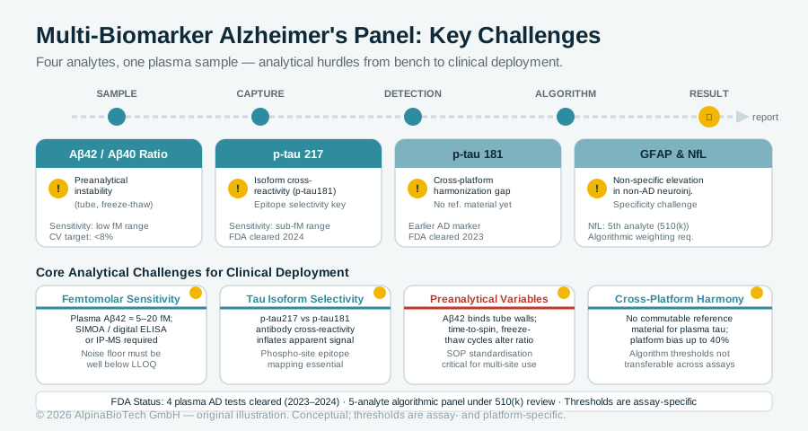
Four Analytes, One Plasma Sample: The Assay Science Behind Multi-Biomarker Alzheimer's Testing
Four blood-based Alzheimer's disease biomarker tests were cleared by the FDA within fifteen months, and a five-analyte algorithmic panel is under 510(k) review. For immunoassay scientists deploying these panels, the analytical challenges of femtomolar sensitivity, cross-reactivity between tau isoforms, preanalytical instability of Aβ42, and cross-platform harmonization are the least-settled problems in making multi-biomarker results hold up outside a research cohort.
Read more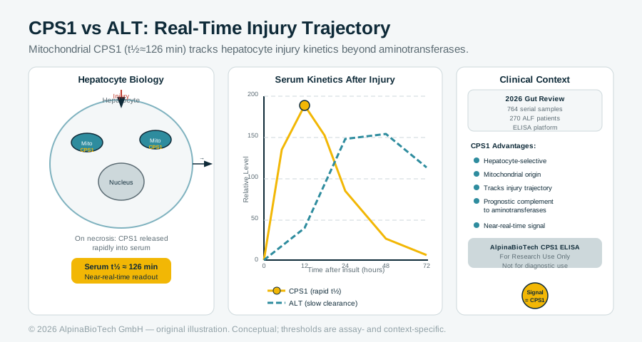
CPS1 Serum Immunoassay: What a Mitochondrial Enzyme Biomarker Tells You That ALT Cannot
Carbamoyl phosphate synthetase 1 (CPS1) is a hepatocyte-selective mitochondrial enzyme with a serum half-life of roughly 126 minutes, making it a near-real-time indicator of injury trajectory rather than injury presence. A 2026 Gut review synthesizes a decade of mechanistic and clinical work, including ELISA data from 764 serial samples across 270 acute liver failure patients, establishing CPS1 as a prognostically informative complement to standard aminotransferase panels. All AlpinaBioTech CPS1 immunoassay products are for Research Use Only.
Read more