Article · 26 June 2026
Understanding the ELISA: Principles, Formats, and Why They Matter for Reproducible Research
The enzyme-linked immunosorbent assay turns a binding event into a measurable signal. Knowing which format you are running is the first step toward data you can trust.
AlpinaBioTech Scientific Team
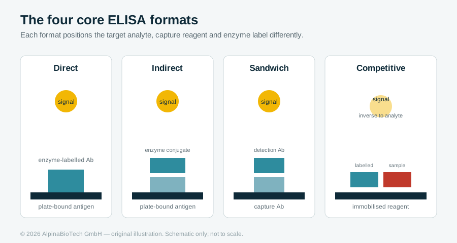
More than fifty years after Engvall and Perlmann first described it, the enzyme-linked immunosorbent assay remains the workhorse of protein quantification in research laboratories. Its appeal is simple: it converts a specific antibody-antigen binding event into a colour change that a plate reader can quantify, without radioactivity and at a scale that suits 96- or 384-well throughput. Behind that simplicity sit several distinct assay architectures, and choosing the wrong one, or running the right one without understanding its limits, is one of the most common sources of irreproducible immunoassay data.
The shared logic
Every ELISA rests on the same three ideas. A capture phase immobilises one binding partner on the well surface. A detection phase introduces an enzyme label, usually horseradish peroxidase or alkaline phosphatase, that is linked to a binding reagent. A substrate is then added; the enzyme converts it to a coloured product, and the optical density is proportional to the amount of label retained in the well. Wash steps between each phase remove unbound material, which is why wash quality so often determines assay quality. The signal is only meaningful relative to a calibration curve built from known standards run on the same plate.
Four formats, four use cases
The direct format labels the primary antibody itself. It is fast and avoids cross-reactivity from secondary reagents, but it is less sensitive and ties up a labelled antibody for every target.
The indirect format adds an enzyme-labelled secondary antibody that recognises the primary. This amplifies signal and lets one labelled secondary serve many primaries, at the cost of an extra incubation and the possibility of secondary-antibody background. Indirect formats are the natural choice when measuring an antibody response, for example, screening serum for antibodies against a defined antigen.
The sandwich format captures the analyte between two antibodies that recognise different epitopes. It is the most specific and sensitive layout for measuring a protein in a complex matrix such as serum, which is why drug-level and many biomarker kits are built this way. Its main constraint is that the target must be large enough to present two non-overlapping epitopes.
The competitive format runs the logic in reverse: sample analyte competes with a labelled reference for a limited number of binding sites, so signal falls as analyte rises. It suits small molecules and haptens that cannot support a sandwich, and it underpins many anti-drug-antibody assays.
Where reproducibility is won or lost
A calibration curve is only as good as its fit. Most quantitative ELISAs are non-linear and are best modelled with a four-parameter logistic curve rather than a straight line forced through a narrow range; reporting results from the flat asymptotes of that curve inflates error dramatically. Matrix effects are the next trap, a result validated in buffer may drift in serum or plasma, so spike-recovery and dilution-linearity checks in the intended matrix are essential. Edge-well evaporation, inconsistent incubation temperature, and pipetting drift across a plate all introduce position effects that a thoughtful plate layout and on-plate controls will expose.
None of these are exotic problems. They are the everyday reasons two laboratories running "the same" assay report different numbers, and they are entirely manageable once the format and its assumptions are understood.
A note on intended use
The kits discussed across these articles are supplied for research use only (RUO). They are powerful tools for method development, translational research, and pharmacokinetic studies, but RUO assays are not validated as in-vitro diagnostic devices and their results should not be used to direct patient care without appropriate independent validation. Treated as research instruments and run with proper controls, well-characterised ELISAs deliver the accurate, reproducible numbers that downstream science depends on.
This article is part of AlpinaBioTech's educational series on immunoassays and therapeutic drug monitoring. For the assay portfolio, see our ELISA kits.
References
-
Engvall E, Perlmann P. Enzyme-linked immunosorbent assay (ELISA). Quantitative assay of immunoglobulin G. Immunochemistry. 1971;8(9):871-874.
-
Lequin RM. Enzyme immunoassay (EIA)/enzyme-linked immunosorbent assay (ELISA). Clin Chem. 2005;51(12):2415-2418.
-
Aydin S. A short history, principles, and types of ELISA, and our laboratory experience with peptide/protein analyses using ELISA. Peptides. 2015;72:4-15.
More articles
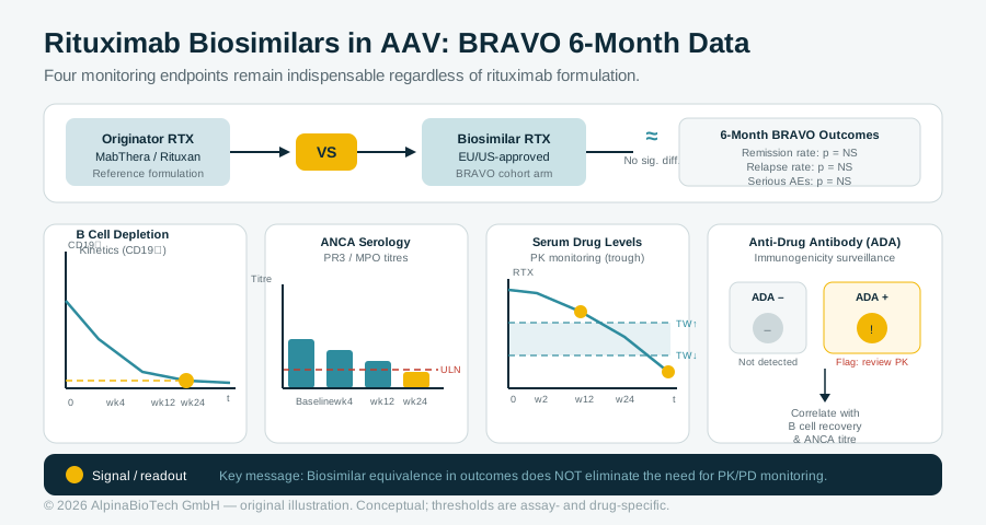
Rituximab Biosimilars in AAV: What Six-Month Real-World Data Tell Monitoring Scientists
The BRAVO multicenter cohort study found no statistically significant differences in remission rates, relapse rates, or serious adverse events between biosimilar and originator rituximab at six months in granulomatosis with polyangiitis and microscopic polyangiitis. For laboratories supporting drug-level and immunogenicity testing in biosimilar-treated AAV populations, the findings reinforce rather than reduce the case for structured pharmacokinetic and pharmacodynamic monitoring. B cell depletion kinetics, ANCA serology, serum drug levels, and anti-drug antibody assays each remain indispensable endpoints regardless of which rituximab formulation the patient receives.
Read more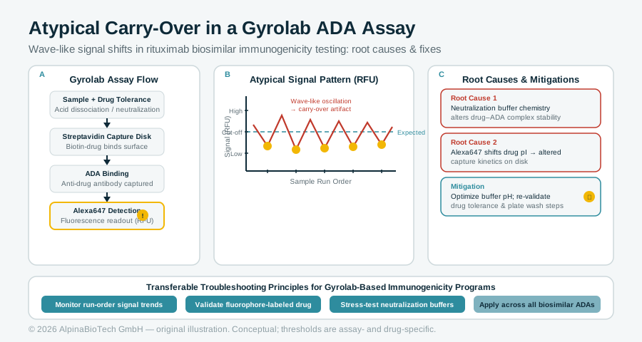
Atypical Carry-Over in a Gyrolab ADA Assay: Lessons from a Rituximab Biosimilar Case Study
A published case study targeting MabionCD20, a rituximab biosimilar, identified wave-like signal fluctuations in Gyrolab-based ADA assays caused by neutralization buffer chemistry and fluorophore-induced isoelectric point shifts. The investigation offers transferable troubleshooting principles for any laboratory running Gyrolab-based immunogenicity programs, particularly around drug tolerance and repeatability.
Read more